PERONEAL TENDON INJURY
(AND OTHER THINGS IN THE LATERAL ANKLE)
There are two tendons on the outside of the ankle, these two work together to turn the foot outward (more specifically - to evert the foot). There is the longer one - called Peroneus Longus, and there is a shorter one called Peroneus Brevis. They live side by side to each other
One of the most common injuries to a body in an inversion ankle sprain (twisting the ankle inward). Then the sprain occurs the ankle twists inward and the tendons and ligaments on the outside of the ankle can get stretched out and torn. There is also an injury that can occur to the cartilage of the ankle when the sprain is severe enough. There are certain people who are more vulnerable to these injuries just because of slight differences in their anatomy ( this is called : anatomical variations and they are normal - there are slight structural differences in all of our bodies ). An extra tendon, a flatter shaped joint, thinner cartilage, thinner ligaments can all contribute to the degree of injury a person can suffer in the long term - from a simple ankle sprain.
There are two kinds of patients when it comes to these injuries of the lateral ankle (outside of the ankle). The person that comes to the office with a sprain that just happened. And the person that comes to our office with an injury that happened weeks or months before. Sometimes they had no pain when it happened, other times they had pain that went away - and then slowly came back.
Lets start with the first patient with the ACUTE injury : the one that just happened. There may be black and blue ( ecchymosis in medical lingo) around the injured area or there may be none- really just depends on the exact nature and degree of damage. We take X-rays of the patients ankle to see if there any injuries to the bone. An x-ray does not show any other structures like ligaments, tendons, or cartilage so in some cases we order an MRI. This kind of test will give us a 3 dimensional view of all the structures inside and if there is an injury an MRI will most likely catch it.
In terms of treatment, in the absence of significant tears or bone damage most people can be treated with casting or a walking boot for a period of time and possibly physical therapy.
A bit more rare of an injury is the tear of the peroneal tendons. This occurs when the ankle turns inward and stretches these tendons against the bones underneath. Many of these tears remain painless but some patients do have persistent pain.
Here are photos and explanations of the most common tendon tears:
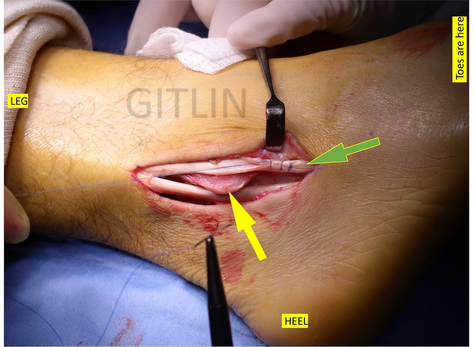
Below is a photograph taken during surgery showing the most common type of peroneal tendon tear. It is usually the peroneus brevis tendon and it is not a tendon tear as you would normally think. Thetendon does not usually just rip in half - although that does happen in trauma cases. This is a “longitudinal tendon tear” meaning the tear in in line with the tendon - its a split lengthwise. The yellow arrow points to the tear, I also labelled where the toes, the heel and the leg are so you can understand the photo better.

The next photo shows a similar tear. The yellow arrow shows the split longitudinal tear, the blue arrow shows a calcification in the tendon ( this usually occurs when there is chronic injury). The green arrow points to a flattened tendon! You will usually think of a tendon as a tubular structure but the peroneus brevis tendon can normally be flat ( long explanation as to why ). In some cases though the tendon does tear and unfold to flatten out. The surgeon needs to make decisions on whether the flat appearance is a type of tear or a normal variant.

The next photo shows the cause of many of these longitudinal tears. The yellow arrow points to a portion of the fibula- the outer ankle bone. The back end of the bone is somewhat sharp and can pierce through the tendon. This is not usually an acute tear and usually happens over time. The green arrow shows the peroneus longus tendon which runs next to and parallel to the peroneus brevis.

The next photo shows a green arrow that points to a peroneus brevis tendon which was found to be flattened from an injury and needs to be fixed. Here we use a technique of tubularization where we suture the tendon in on itself and make it a round structure. This is not done with every case.
Another photo below showing a completely repaired tendon using the tubularization technique.

There is a lot more to injury of these tendons, the surgeon needs to assess muscle bulk, bone tunnel depths, and ligament injury to the area. Many times the tendon is not the only structure that needs to be repaired, there other structures need to be addressed to prevent later damage to the tendon.