CALCANEAL (HEEL BONE) FRACTURE
WHAT IS A CALCANEAL FRACTURE ?
This is a broken heel bone, most times the patient falls from a height like a ladder or simply missed a stair or two and falls onto their one foot. This bone is normally very strong but when all the body weight comes crashing down on the small surfaces of the joint it is easy to crush the bone. These are very serious injuries since when the bone is crushed the damage to the subtalar joint is severe and many times can lead to disfigurement of the foot and arthritis and tendon issues.
HOW ARE CALCANEAL FRACTURES DIAGNOSED AND ASSESSED?
Generally we can get most information from a series of foot and ankle X-rays but in certain cases we may order a CAT scan. This will give us a 3D and interior view of the heel bone and show us all the fragments and the extent of the injuries. Depending on the amount of injury to the bone we would then consider whether surgery is warranted.
IS SURGERY ALWAYS NECESSARY TO FIX A HEEL FRACTURE?
Surgery for a broken calcaneus heel bone is not always needed. If the fragments are so displaced that the bone does not resemble its normal anatomy then yes surgery may be necessary. especially when those fragments make up the subtalar joint. This is an important joint in the back of the foot that allows us to move the foot from side to side. If the bone loses its shape surgery may be necessary.
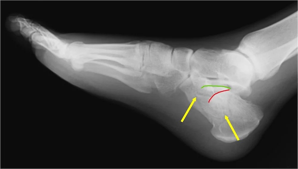
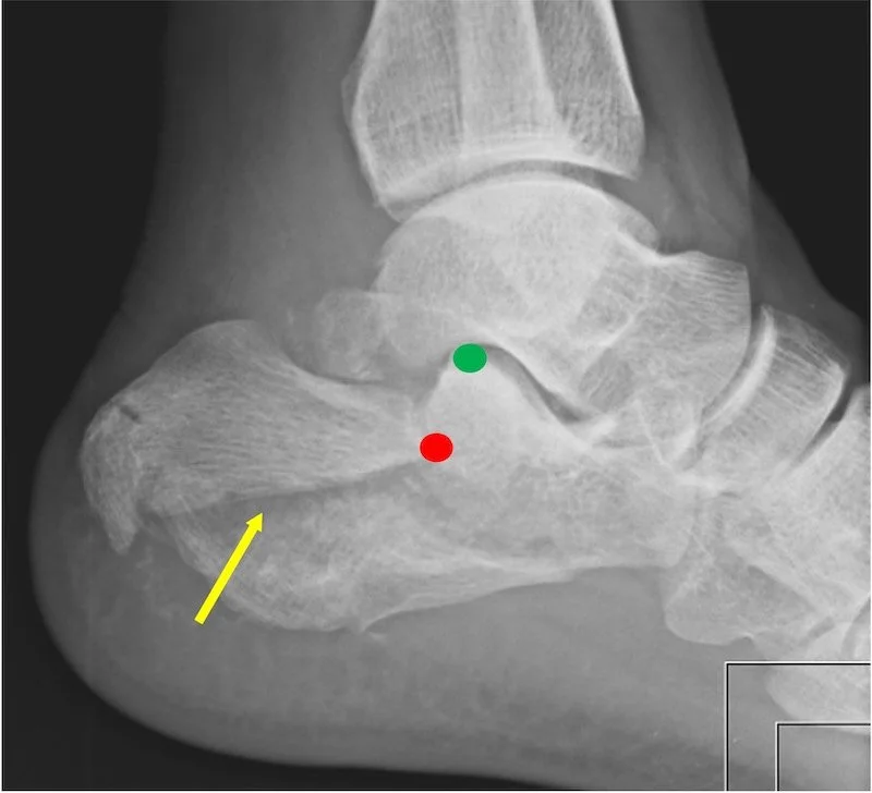
The first picture is what is called a joint depression fracture. the body weight lands hard on the foot and crushes the joint pushing the cartilage down into the actual interior of the heel bone. The yellow arrows show fracture lines, there are many fracture peices in this case. The red curved line shows the position of the subtalar joint that is displaced downward, forced there by the body weight. The green curved line shows where it used to be before is was crushed down.
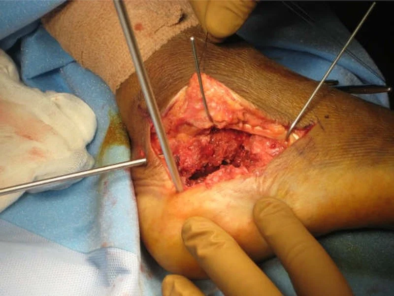
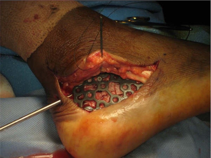
You can see in this picture during surgery that the bone has a black dark area through it , thats how the fracture line looks. When these bones break the bone is crushed into itself and we essentially lose bone shape. During surgery we put it all together as much as possible. Then we apply a large plate that you can see in the next photo tp hold all of the fragments together. As you can see this is a large incision, we prefer not to do it this way so 90 percent of the time we are able to use or minimal incision technique for these bone repairs.
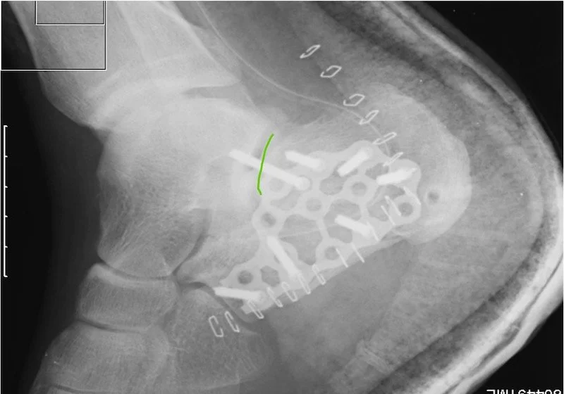
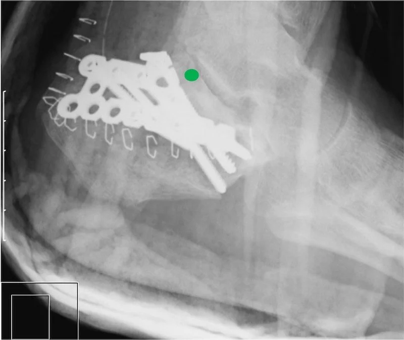
Here is an xray of the same foot with a plate attached after surgery to realign the joint and bones. You can see the curved green line- we were able to put the subtalar joint back into alignment to hopefully prevent progression of arthritis and pain.
WHAT KINDS OF CALCANEAL FRACTURE PATTERNS AREA THERE?
We generally divide the fractures into two categories, above we showed a joint depression fracture, here below is the tongue type fracture. It is called that because the fracture fragment “looks like a tongue”. The pattern is different because the position of the foot was different when the body weight came down into the foot. The red dot shows the front edge of the tongue fracture and that red dot should actually be where the green dot is. The yellow arrow points to the fracture line which is easy to see in this case. The next xray shows only the green dot because we repaired the fracture and realigned it. You cn see the surgical plate holding the bone together.
IF YOU NEED HELP OR ADVICE PLEASE EMAIL US BELOW AND WE WILL GET BACK TO YOU AS SOON AS POSSIBLE
Our Florida and New York Office Locations
NAPLES OFFICE (Gulf Coast)
Serving Southwest Florida, including Fort Myers, Cape Coral, Bonita Springs, and Sarasota.
Address: 3940 Radio Road, Unit 104, Naples, FL 34104
Phone: 239-465-0311
WESTON OFFICE (Atlantic)
Serving the Tri-County area, including Miami, Boca Raton, West Palm Beach, and Hollywood.
Address: Weston near Cleveland Clinic coming soon
Phone: 239-465-0311
NEW YORK CITY OFFICE
Serving New York City and the boroughs as well as the entire Tri-state area
Address: 330 W. 58 street, Unit 610, NYC, NY 10019
Phone: 212-372-0991